Bi2S3-SiO2 NRs/二氧化硅涂层硫化铋纳米棒多模态显影剂可用于胃肠道GI成像技术
利用硫酸钡悬浮液作为X光显影剂用于小肠成像,由于其自身具备的非降解及其他一些不理想的特点使其在胃肠道穿孔及肠道结构观测方面受到很大限制;其他常用的胃肠道显影剂便是碘代分子,但因其本身X射线吸收率较低,所以通常需要大剂量使用才能达到理想的效果,这常常会使病人产生碘过敏性反应。
制备得到二氧化硅涂覆的硫化铋纳米棒(Bi2S3@SiO2 NRs)作为多模态显影剂用于胃肠道的无侵害性实时成像以及直接观测其在胃肠道下部的流经过程(Scheme 1)。经二氧化硅包覆后,在胃及小肠中,Bi2S3@SiO2 NRs展现出非常好的水溶性,生物相容性以及稳定性。通过TEM可以看出,Bi2S3 NRs宽约10 nm,长约50 nm,元素分析谱图显示成功制备得到高纯度的Bi2S3 NRs(Fig. 1),经二氧化硅涂覆后,Bi2S3@SiO2 NRs呈现单分散性,SiO2壳层厚度约为6 nm(Fig. 2)。
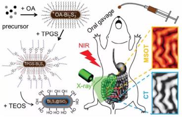
Scheme 1 Schematic illustration of the preparation of Bi2S3@SiO2 NRs for bimodal CT/PAT imaging of the GI tract principle based on the unique properties of Bi2S3@SiO2 NRs。
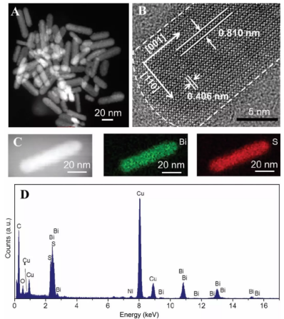
Fig. 1 Characterization of Bi2S3 NRs. (A) HAADF-STEM image and (B) HRTEM image of Bi2S3 NRs prepared by the solvothermal method. (C) Corresponding element mapping for Bi and S of the as-prepared Bi2S3 NRs. (D) EDS of the as-prepared Bi2S3 NRs.
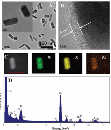
Fig. 2 Characterization of Bi2S3@SiO2 NRs. (A) TEM image and (B) HRTEM image of as-prepared Bi2S3@SiO2 NRs. Inset: HAADF-STEM image of Bi2S3@SiO2 NRs. (C) Corresponding element mapping for Bi, S and Si of Bi2S3@SiO2 NRs. (D) EDS of Bi2S3@SiO2 NRs.
通过CT成像分析发现,随着Bi2S3@SiO2 NRs浓度增加,其HU值明显增加,相同浓度下的HU值明显高于硫酸钡,PAT信号随浓度增加也呈线性增长关系(Fig. 3)。
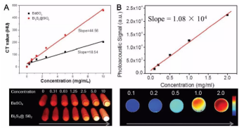
Fig. 3 CT and PAT phantom images of Bi2S3@SiO2 NRs with different concentrations in vitro. (A) Plot of Hounsfield units (HU) values and of Bi2S3@SiO2 NRs and BaSO4 suspension versus the sample concentrations and CT phantom images of Bi2S3@SiO2 NRs and BaSO4 suspension samples with different concentrations. (B) Plot of the photoacoustic signal versus Bi2S3@SiO2 NRs concentrations and PAT phantom images of Bi2S3@SiO2 NRs aqueous solutions with different concentrations.
对Bi2S3@SiO2 NRs的生物相容性进行检测,发现16HBE与Bi2S3@SiO2 NRs共培养24 h后并未显示出明显的毒性,并且该粒子进入秀丽隐杆线虫体内后对其寿命也没有明显的影响,这就说明Bi2S3@SiO2 NRs具有非常好的生物相容性(Fig. 4)。
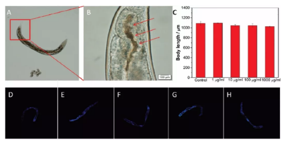
Fig. 4 Biosafety assessment of Bi2S3@SiO2 NRs by the C. Elegans model. (A) Bright field image of the NRs distribution in the GI tract of C. Elegans. Worms feed on NGM plates with Bi2S3@SiO2 NRs (1000 μg mL−1) transferred onto an agar pad after 1 h. (B) The distribution of food containing Bi2S3@SiO2 NRs (red arrows) in the intestine of the worm’s tail. (C) Effects of Bi2S3@SiO2 NRs with different concentrations on body length of C. Elegans. (D–H) Effects of Bi2S3@SiO2 NRs treatments on the accumulation of lipofuscin in age-synchronized worms. Representative fluorescent images of worms fed with 0, 1, 10, 100 and 1000 μg mL−1 Bi2S3@SiO2 NRs, respectively.
Bi2S3@SiO2 NRs以口服的方式进入BALB/c裸鼠体内,通过CT及PAT对Bi2S3@SiO2 NRs进行实时成像追踪,发现该粒子通过胃肠道渐渐进入小肠较后以粪便的形式排出体外,该过程中对胃肠道的功能没有明显的损伤,说明Bi2S3@SiO2 NRs对组织无侵入性损伤(Fig. 5, 6, 7)。
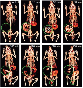
Fig. 5 CT imaging of the GI tract in vivo. In vivo X-ray CT imaging of the GI tractin BALB/c nude mice at different intervals after oral administration of Bi2S3@SiO2 NRs.
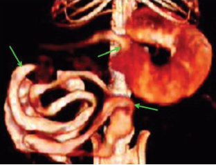
Fig. 6 Enlarged images of CT images of the GI tract of mice 30 min post oral administration of Bi2S3@SiO2 NRs.
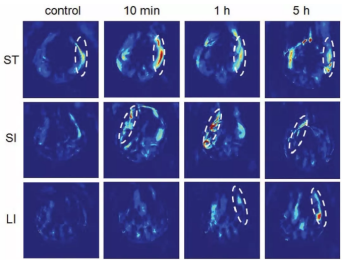
Fig. 7 PAT imaging of the GI tract in vivo. PAT cross-sectional image of the GI tract of BALB/c nude mice at different intervals after oral administration of Bi2S3@SiO2 NRs: stomach (ST), small intestine (SI) and large intestine
wyf 04.02


 齐岳微信公众号
 官方微信
官方微信 库存查询
库存查询