文献:Exosomal Vaccine Loading T Cell Epitope Peptides of SARS-CoV-2 Induces Robust CD8+ T Cell Response in HLA-A Transgenic Mice
作者:An-Ran Shen 1, Xiao-Xiao Jin 2, Tao-Tao Tang 1, Yan Ding 2, Xiao-Tao Liu 2, Xin Zhong 1, Yan-Dan Wu 2, Xue-Lian Han 3, Guang-Yu Zhao 3, Chuan-Lai Shen 2, Lin-Li Lv 1,✉, Bi-Cheng Liu 1
文献链接:https://pmc.ncbi.nlm.nih.gov/articles/PMC9346304/
摘要:
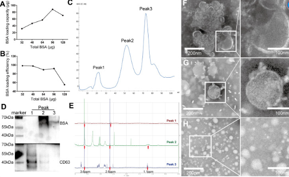
The feasibility of this novel approach in generating peptides-PEG-lipid-exosome complexes was first tested by preparing BSA-PEG-lipid-exosome complexes. The molar ratio between BSA and DPME-PEG-NHS was optimized by testing 1:1, 1:1.5, 1:3, and 1:4 ratios as shown in Table 1. In order to calculate the BSA loading capacity and loading efficiency in the BSA-PEG-micelles, the conjugated BSA with DMPE in BSA-PEG micelle solution and the free BSA in the filtered solution was quantified using BCA kit, respectively. The results showed that the BSA loading capacity was highest (85 μg BSA was conjugated with 2 mg DMPE-PEG-NHS) at 1:3 molar ratio of DMPE-PEG-NHS with BSA (Figure 3A). The loading efficiency was 91.84% at 1:3 molar ratio and then decreased rapidly when the BSA input increased (Figure 3B). Then, the RBC-exosomes were mixed with BSA-PEG-micelles in a 1:1 ratio based on the protein concentrations. The mixture was incubated in a water bath for 2 h at 40℃. The BSA-PEG-lipid-exosomes solution was then cooled to 4℃ and instantly purified using size-exclusion chromatography.

通过制备BSA-PEG脂质外泌体复合物,首次测试了这种新方法产生肽-PEG-脂质外泌物复合物的可行性。如表1所示,通过测试1:1、1:1.5、1:3和1:4的比例,优化了BSA和DPME-PEG-NHS之间的摩尔比。
为了计算BSA-PEG胶束中的BSA负载能力和负载效率,分别使用BCA试剂盒定量BSA-PEG胶束溶液中与DMPE偶联的BSA和过滤溶液中的游离BSA。
结果表明,当DMPE-PEG-NHS与BSA的摩尔比为1:3时,BSA的负载能力最高(85μg BSA与2mg DMPE-PEG-NaHS偶联)(图3A)。在1:3摩尔比下,负载效率为91.84%,然后随着BSA输入的增加而迅速降低。
然后,根据蛋白质浓度,将RBC外泌体与BSA-PEG胶束以1:1的比例混合。将混合物在40℃的水浴中孵育2小时。然后将BSA-PEG脂质外泌体溶液冷却至4℃,并立即使用尺寸排阻色谱法纯化。
BSA-PEG脂质外泌体的纯化与鉴定。
首先将一系列量的BSA与2mg DMPE-PEG-NHS偶联,然后与DSPE-PEG混合形成BSA-PEG胶束。对游离BSA进行定量,并分别计算BSA-PEG胶束中的BSA负载能力(A)和负载效率(B)。
此外,BSA-PEG胶束以DMPE-PEG-NHS与BSA的1:3摩尔比再生,并在40℃下与RBC外泌体一起孵育2小时,以产生BSA-PEG脂质外泌体,并使用尺寸排阻色谱法立即纯化,FPLC图谱显示三个主峰(C)。
用抗BSA和抗CD63对三个峰的组分进行蛋白质印迹,以鉴定每个峰中的BSA和外泌体(D),并对每个峰中PEG、水解NHS和DMPE元素进行液体NMR鉴定(E)。
相关推荐:
DSPE-Poly(L-lysine)
DSPE-Pyrene
DSPE-Cap-Biotin
DSPE-Cap-Folate
DSPE-PEG-OH
DSPE-PEG-HZ
DSPE-PEG-Mal
DSPE-PEG-N3
以上文章内容来源各类期刊或文献,如有侵权请联系我们删除!


 齐岳微信公众号
 官方微信
官方微信 库存查询
库存查询